Academics from the School of Computer Science and Engineering won the Best Paper Award at the IEEE International Symposium of Computer-Based Medical Systems for their research paper titled ‘3D Deep Learning for Anatomical Structure Segmentation in Multiple Imaging Modalities’.
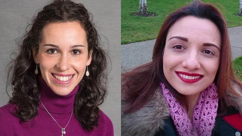
Dr Barbara Villarini, Lecturer in Software Engineering, and Dr Hykoush Asaturyan, Researcher in Medical Image Computing, presented their paper at the symposium virtually, which describes how they used 3D deep learning models to extract information about the pancreas, liver, kidneys and iliopsoas muscles from adults and children using both MRI and CT scans. The academics established a clinical-based and technical collaboration with Harvard Medical School and Boston Children’s Hospital, and their work hopes to enable disease progression to be classified and monitored.
Their study focused on the development of a novel computer program that extracts anatomical structures of organs and muscles in 3D MRI and CT scans by harnessing the advantages of deep learning technology in artificial intelligence (AI). This work addresses the critical technical challenges related to high variations in anatomical structure and size, varying image resolutions from MRI and CT scanners and blurring in a patient’s resultant scans.
The team developed an AI-driven computer program that combines two different neural networks using 3D deep learning technology. The first network outlines a bounding box around the target organ or muscle in a scan, and the second performs accurate segmentation in the region localised by the bounding box. This was successfully evaluated on six different datasets, including MRI, DCE-MRI and CT scans targeting the pancreas, liver, kidneys and iliopsoas muscles.
There is a consistent rise in the number of medical diagnostic imaging tests, with over 3 million MRI scans and over 5 million CT scans taking place annually nationwide, which take a long time to analyse to assess treatment plans and can include laborious manual segmentation. The researchers’ computer program can accelerate pre-clinical investigations and reduce radiologists’ time and burden, eliminating or minimising the need for manual segmentation.
Dr Villarini, Dr Asaturyan and their team hope that their novel computer program could be deployed into a computer-aided system to support biomedical research. They said that through the system, it may also be possible to investigate further correlations between significant changes in liver volume and alcoholic liver disease, kidney volume and polycystic disease, or between the volume of psoas muscle and predictive outcomes of treatment for bladder cancer chemotherapy and ovarian cancer.
The paper is part of a longstanding collaboration between Professor Jimmy Bell and Professor Louise Thomas from the Research Centre for Optimal Health at the University of Westminster alongside Dr Villarini and Dr Asaturyan from the School of Computer Science and Engineering, and it was supported by the Royal Academy of Engineering Leverhulme Trust Fellowship, recently awarded to Dr Villarini.
Talking about the award, Dr Barbara Villarini said: “I am delighted to have received the ‘Best Paper Award’ in the 34th IEEE Symposium on Computer-Based Medical Systems (CBMS). Our paper has been selected among more than 70 presented works and this is a high honour. I am very proud of our team and the work that we are carrying out.”
Dr Hykoush Asaturyan added: “I am so grateful for the interdisciplinary and multidisciplinary contributions of our entire team and look forward to continuing our collaboration. It is with delight to learn that our paper was awarded first prize in the 34th IEEE Symposium on CBMS, one of the leading conferences within the fields of medical and biomedical informatics.”
Related articles


